Ophthalmology
When I've got a new job at a practice of an ophthalmologist (in this one), there was a lot of opportunity to learn a lot of new fascinating areas of the human body. In this case it was the human eye, in which I was fascinated long before I even started to work. I think of myself to be an "eye person", because when I meet someone I first look into his or her eyes. I think the saying: "The eyes are a window to the soul" is not so far from the truth as one might think. So my new job couldn't be more fitting.
I was impressed by the many diagnostics and ways to treat conditions of the eye. Therefore, I had to draw most of them of course.
DIL
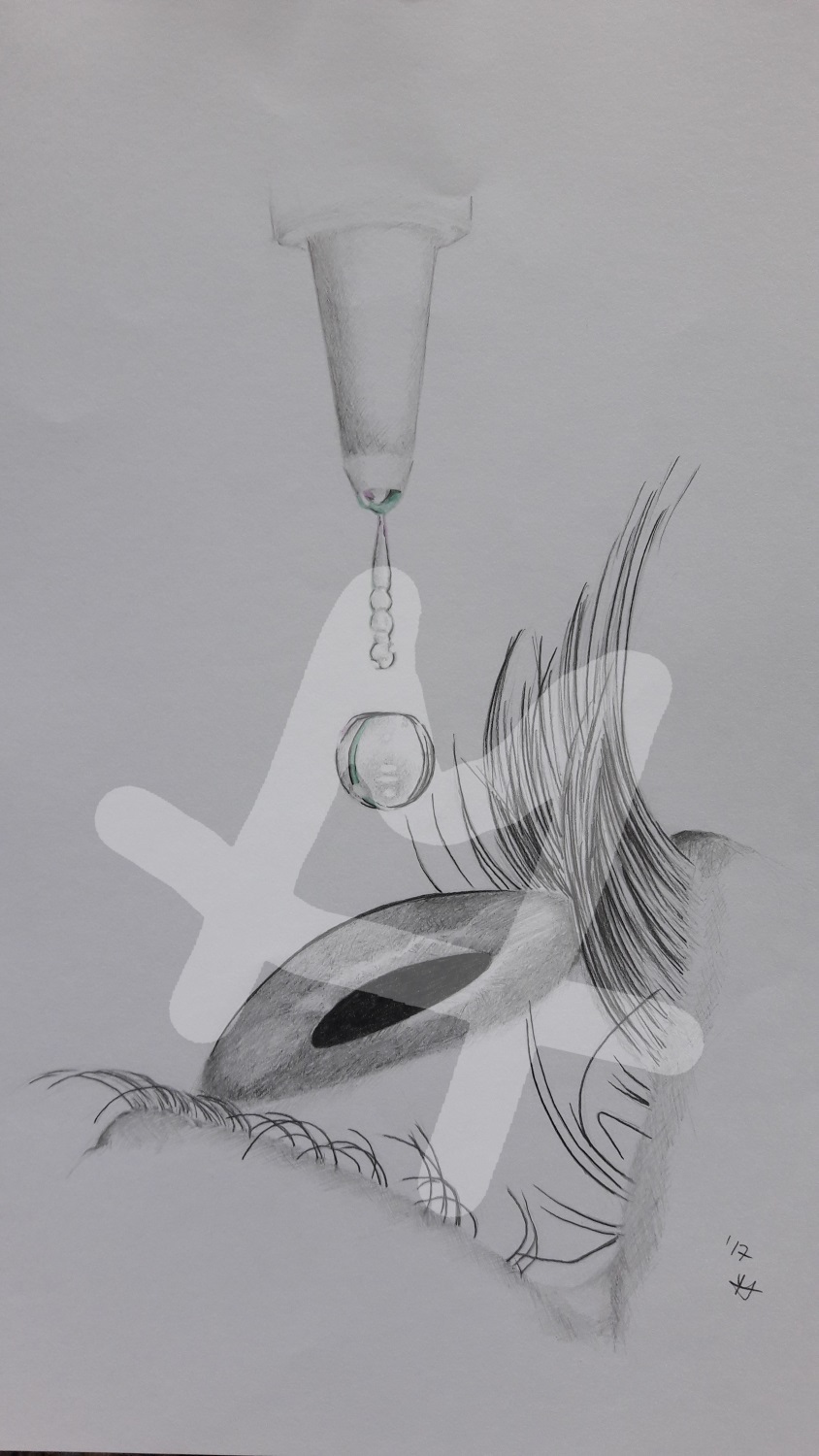 To be able to examine the eyes properly it's often necessary to enlarge the pupils with special eye drops. This dilatation (short DIL) will widen the pupils so an ophthalmologist can look inside the eye. The great disadvantage of this is a prohibition to drive a car for at least four hours. With dilated pupils the eye isn't capable of seeing sharp.
OCT
With the Optical Coherence Tomography (OCT) it's possible to map all the distinctive layers of the retina. So an ophthalmologist can see the macula, the area of the clearest vision or the optic nerve itself.
When there's the suspicion of an age-related maculare degeneration (AMD) a Macula-OCT is necessary, to check for possible fluid leakage. At the beginning the pupils are dilated with eye drops to make it easier to examine the retina, because more light is getting into the eye. So at the end there won't only be a scan of the retina but a foto of the retina itself. If the OCT is done without dilated pupils the retina can't be fotographed, because not enough light can get through.
The layers of the retina are mapped with a harmless laser which is reflected by the thickness of each layer differently.
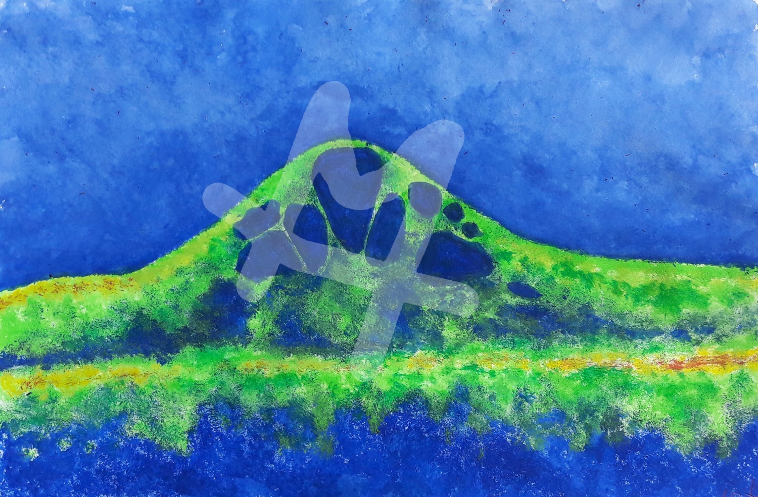
If there's a diabetic macular edema (s. picture) it's shown by a lot of cysts filled with fluid in the area of the macula. This can affect the vision in many ways.
FA
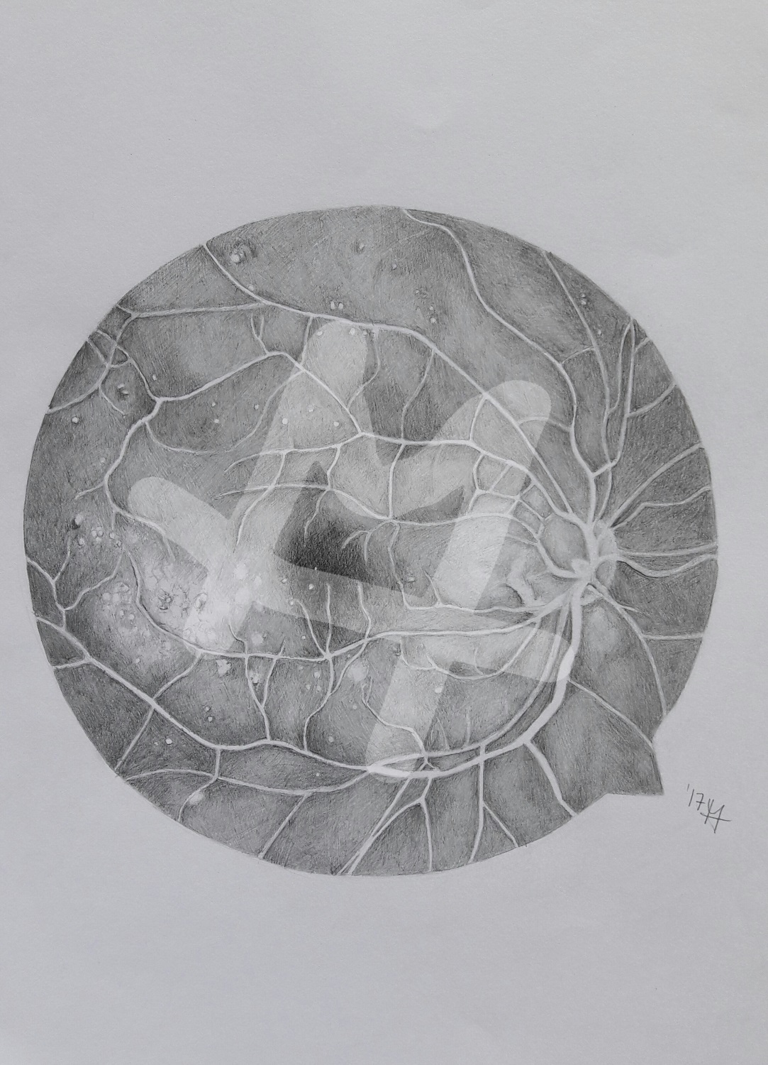 With a Fluorescence-Angiography (FA) it's possible to look at the blood vessels of the retina. Right after the pupils were dilated a fluorescein is injected intravenously. An interesting side effect of this solution is that the patient now has a colorful vision (e.g. if the fluorescein is red the vision appears pink). When the solution is injected a serious of picture is taken in a short period of time. So the ophthalmologist is able to see the filling of the blood vessels to look after abnormal or leaking ones. To have a comparison always both eyes are examined. But the important one gets all the attention escpecially during the injection phase. After a couple of minutes another series of pictures is taken and short after that another. In that way it's possible to look for abnormalities.
Cataract surgery
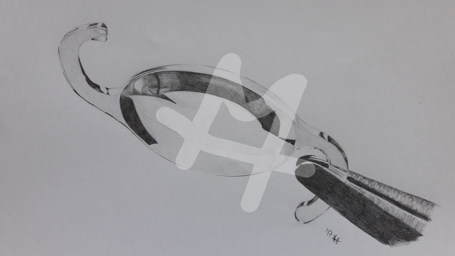 The, mostly due to old age, cataract is an opacification of the natural lens of the eye - a clouding of the vision so to speak. Therefore, it's harder to differentiate sharp forms, colors or contrast. It's not possible to treat the cataract with medicine yet and so a surgery has to be performed in which the natural lens is replaced by an artificial one. The necessary intraocular lens (IOL) requires special measurements to guarantee a better vision after the surgery. That would be the acuteness of vision, refraction power and different measurements of the eye itself, e.g. the depth of its anterior chamber. But before surgery other disseases must be excluded.
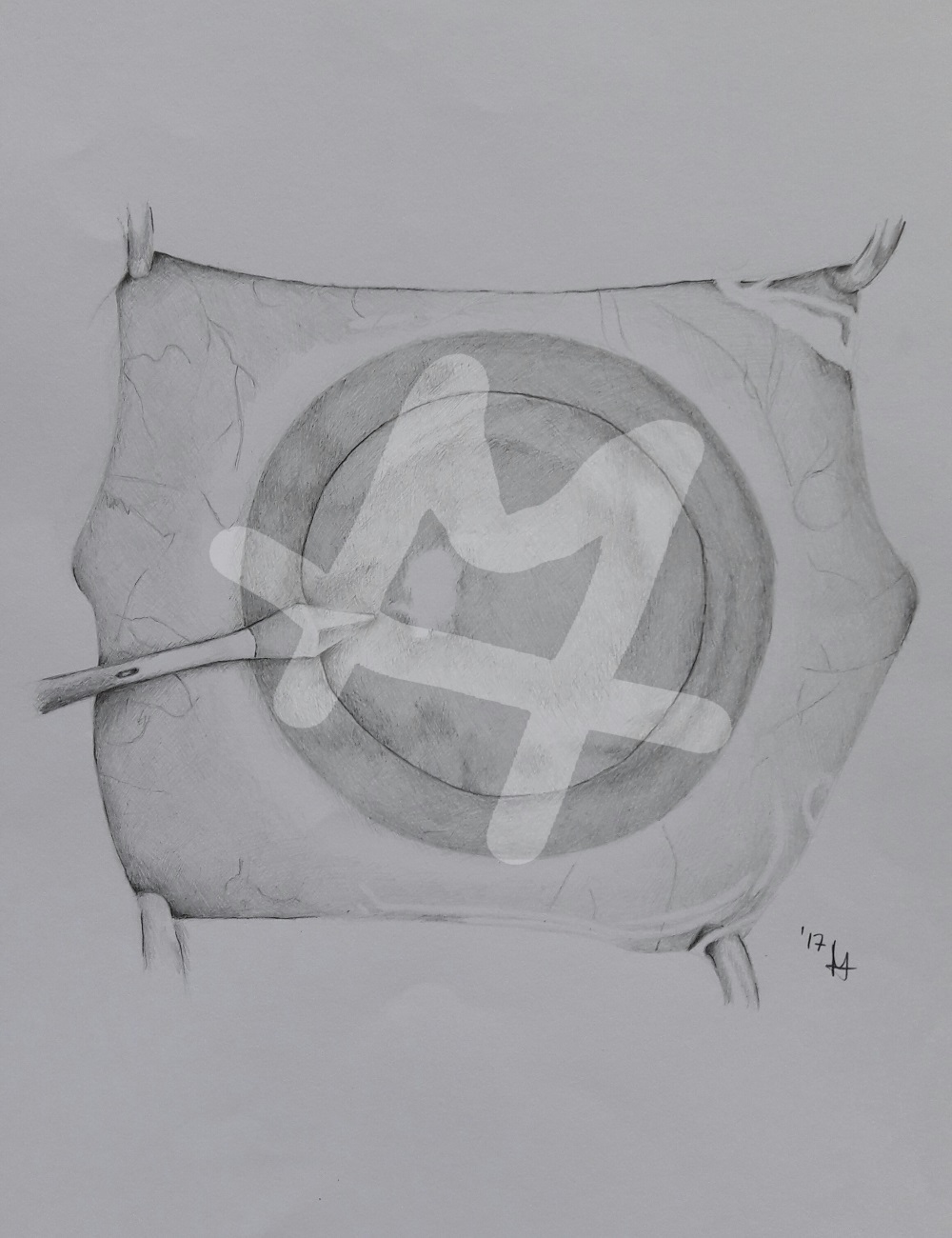 Before surgery is starting the pupil is enlarged with eye drops. After preparing the area three small incisions are made in the cornea to get access to the lens capsule. Now a viscoelastic fluid is injected to the front part of the eye to get more space in the anterior chamber of the eye and to keep its shape. Also it's protecting the outer layer of the cornea and covers the instruments used during surgery.
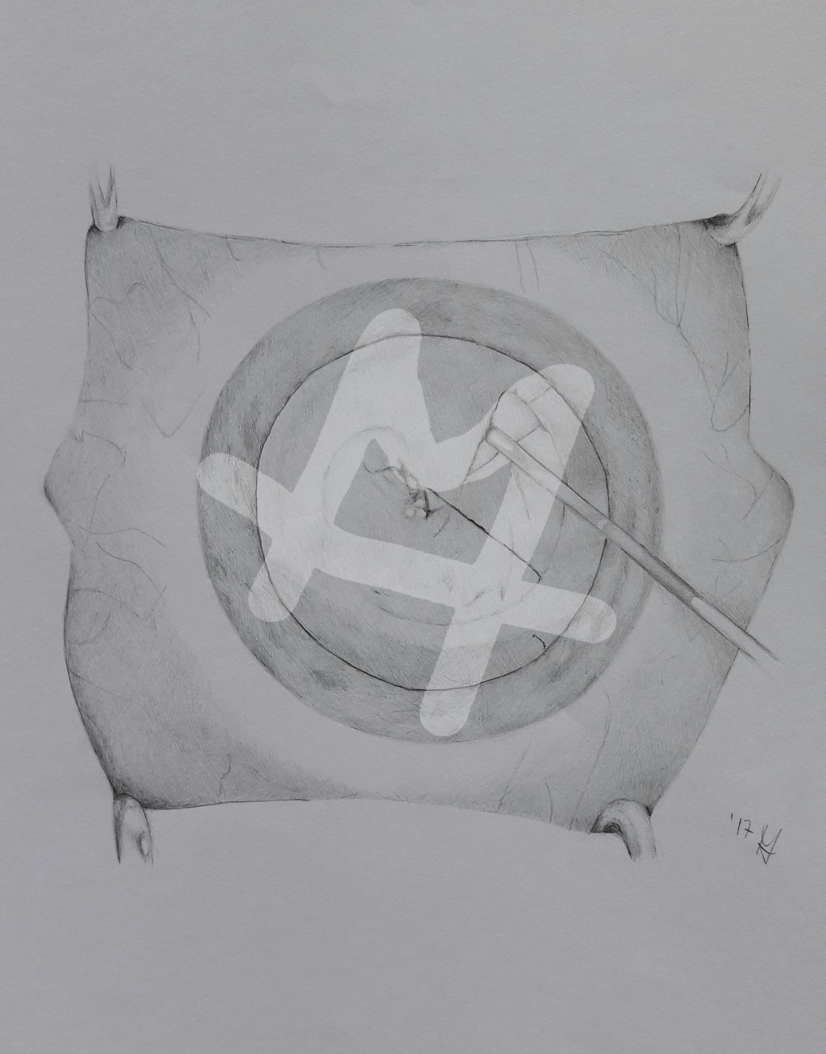 With a special instrument the surgeon performs a so called capsulorhexis, in which the anterior capsule of the lens is scratched circularly and opened.
After that the lens is untightend from the lens capsule with a water filled cannula (Hydrodisection).
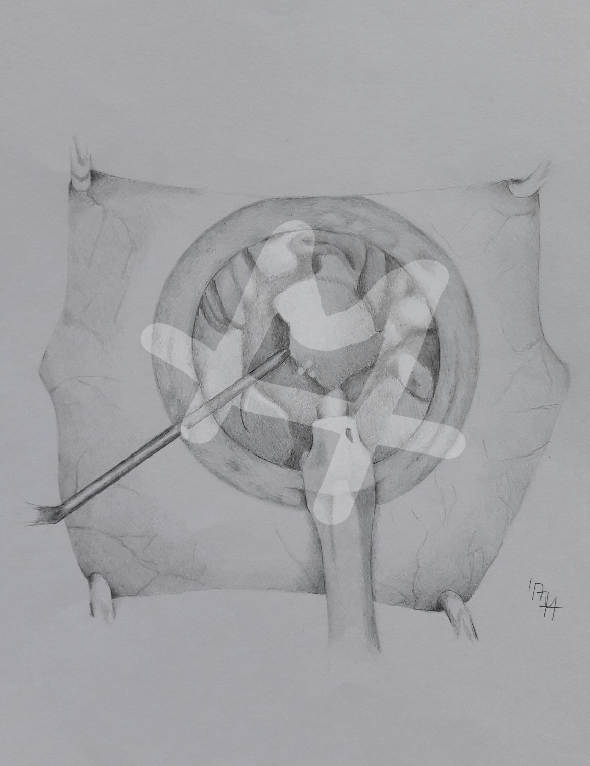 By using a hollow needle and a special ultrasound device the lens gets crushed and sucked off. During that procedure the surgeon gently turns the lens with the hollow needle. This is also called Phacoemulsification.
After the rough removal of the lens the remaining pieces are removed by a device which can irrigate and aspirate. Now the artificial lens is placed into a cartridge where it gets fold automatically, so it fits through one of the small incisions in the cornea.
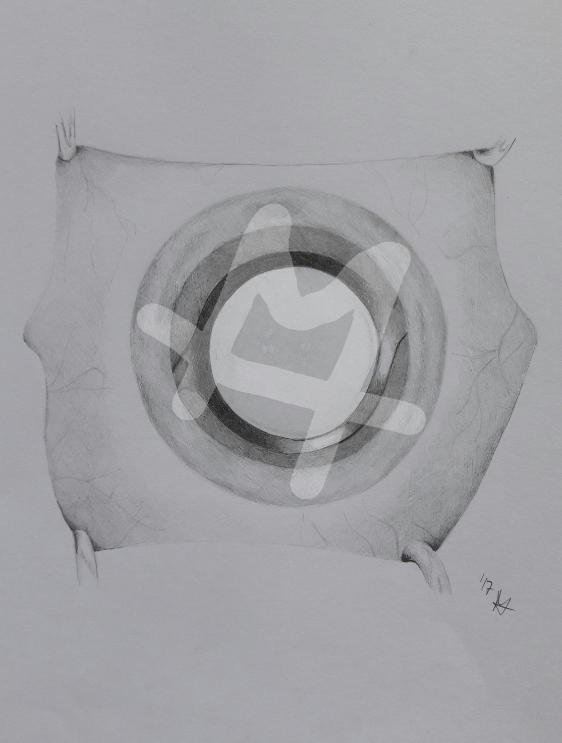 Before the actual implantation a viscoelastic fluid is injected one more time.
Then the cannula is carefully inserted into the lens capsule where the folded lens slowly unfolds. After the viscoelastic fluid is sucked off under and above the artificial lens it can be put into its right place.
The surgery is performed ambulant so the patient can go home short after the procedure. He has to take anti-inflammatory eye drops and has to see the ophthalmologist the next day. A few days later another checkup is necessary to make sure everything went well.

 Dies ist eine mit page4 erstellte kostenlose Webseite. Gestalte deine Eigene auf www.page4.com